


Introduction
Traditional anatomy education, built on textbooks, plastic models, and cadaveric dissections, faces significant challenges in today’s classrooms. As medical and biology programs expand and technology advances, educators and students are increasingly turning to 3D printing as an innovative solution to bridge gaps in knowledge, engagement, and accessibility.
3D printing in anatomy education offers a powerful way to create realistic, affordable, and customizable anatomical models. With technologies like FDM 3D printing, SLA printing, and additive manufacturing, institutions can enhance spatial understanding, boost student engagement, and make learning more interactive.
Common Issues in Traditional Anatomy Education
1. Limited Access & Cost
Cadaveric dissection is often costly and logistically challenging. Many schools struggle to maintain enough cadaver specimens or physical models for all students, impeding hands-on learning.
2. Poor Spatial Understanding
2D images in textbooks cannot capture the complexity of anatomical structures. Students often struggle to visualize the real relationships between organs, nerves, and bones, which leads to knowledge gaps and poor exam performance.
3. Lack of Engagement
Passive learning from lectures or static models fails to excite or motivate students, resulting in low retention, poor practical skills, and disengagement.
4. Sanitary and Ethical Concerns
Working with human specimens can cause discomfort or ethical concerns for some learners. Fixed tissue poses hazards, while accessing fetal or pathology samples is nearly impossible for most institutions.
5. Assessment Limitations
Traditional methods make it hard to assess complex practical skills or accommodate individualized learning paths.
How 3D Printing Solves Key Challenges
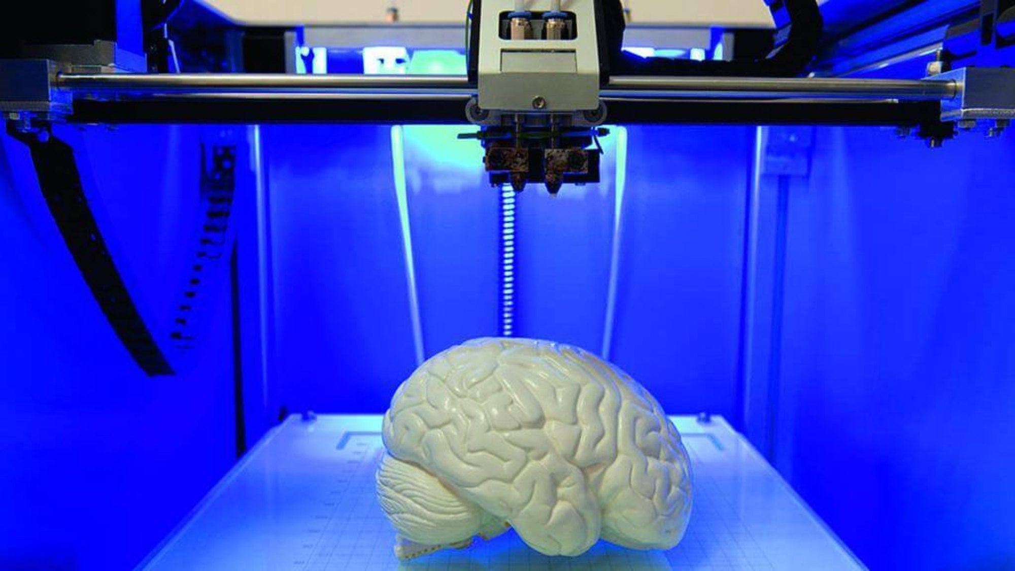
3D printing, also known as additive manufacturing, produces highly accurate and customizable models for anatomy education. These models can be designed and developed using advanced tools such as Paint 3D for educational purposes, and printed using various technologies like FDM (Fused Deposition Modeling) or SLA (Stereolithography).
1. Improved Spatial Understanding
- Tactile, Manipulable Models: Students can touch, rotate, and explore anatomical models, gaining a clearer sense of structure and orientation than with flat images.
- Visualization of Complex Structures: Neuroanatomy and intricate systems become easier to learn, reducing confusion and building expertise in fields like surgery and radiology.
2. Enhanced Engagement and Learning
- Active, Hands-on Approach: Practical interaction with 3D-printed models creates immersive lessons that boost retention and satisfaction.
- Student Motivation: Medical students demonstrate greater interest and participation when training with realistic models—leading to better exam results and skills.
3. Accessibility and Cost-Effectiveness
- Affordable Production: 3D printing brings down costs, allowing even remote or budget-constrained institutions to generate high-quality models.
- Broad Reach: Anyone with a 3D printer and digital resources can benefit—expanding access to anatomy education worldwide.
4. Realistic Representations
- True-to-life Models: Modern product development workflows replicate not only normal anatomy but also pathologies, rare variations, and surgical scenarios.
- Customized Solutions: Models can be tailored to curriculum needs, specific cases, or particular student groups.
5. Flexibility and Customization
- Rapid Prototyping: New anatomical models can be designed, previewed, and printed quickly using additive manufacturing techniques.
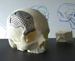
- Patient-Specific Teaching Tools: Unique organs, bones, or pathological structures can be printed from real patient scans.
6. Reduced Reliance on Cadavers
- Less Need for Human Specimens: Ethical, cultural, and legal issues are alleviated, and students uncomfortable with dissection can still learn effectively.
- Expanded Learning Scenarios: Rare or difficult-to-sample specimens become readily accessible.
7. Improved Assessment
- Objective Skill Testing: 3D-printed models help evaluate surgical, diagnostic, and anatomical skills with precision.
- Spiral Curriculum Support: Models help scaffold knowledge from basics to advanced anatomy.
3D Printing Applications and Case Examples
- Undergraduate Medical Classes: 3D-printed models boost performance in anatomy assessments, especially in learning cardiac, skeletal, and neuroanatomical structures.
- Surgical Training: Students perform better using printed tissue and organ models, leading to increased technical skills and interest in surgery.
- Pathology Education: Models of rare disease states help students understand cases they’d otherwise never see.
- Remote/Resource-poor Settings: Rural clinics and international schools can print their anatomical resources locally.
Step-by-Step: Bringing 3D Printing to Anatomy Class
1. Digital Modeling
Use tools like Paint 3D or medical imaging data to design custom anatomical models.
2. Choosing the Right Printing Technology
- FDM 3D Printing: Uses heated filament to build sturdy, cost-effective models.
- SLA Printing: Uses photopolymerization to create high-resolution, detailed models.
3. Material Selection
Choose PLA or ABS filament for FDM, or resin for SLA printing.
4. Printing and Finishing
Print models in-house or via service bureaus, then paint and finish for realism.
5. Classroom Integration
Replace or supplement cadaveric study with printed models, design practical assessments, and encourage group interaction.
Evidence Supporting 3D Printing in Anatomy Education
- Comparative Studies: Better academic performance and skills when 3D-printed models replace or supplement traditional methods.
- Surveys: Students and educators strongly support integrating printing technologies into medical curricula.
- Practical Trials: Suture training, manipulation, and exam results show strong improvements.
Limitations and Considerations
- Fidelity vs. Cost: Models may not perfectly replicate texture or color of real tissue, though new materials are closing the gap.
- Maintenance and Expertise: Integration requires digital modeling skills and printer upkeep.
Conclusion: Unlocking Tomorrow’s 3D Printing Anatomy Education Today
By harnessing the power of 3D printing, educational institutions can create realistic, affordable, and customizable anatomical models using FDM 3D printing, SLA printing, and modern product development techniques.
This revolution closes gaps in spatial understanding, engagement, and accessibility—making learning more vivid, motivational, and effective for students everywhere. Whether you’re an educator modernizing your curriculum or a student seeking deeper understanding, adopting additive printing and 3D designing is the key to mastering the human body in the 21st century.
